THE BIOTECHNOLOGY
EDUCATION COMPANY
EDUCATION COMPANY
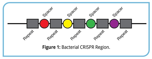 In 1987 Yoshizumi Ishino and colleagues at Osaka University in Japan were researching a new microbial gene when they discovered an area within it that contained five identical segments of DNA made up of the same 29 base pairs. The segments were separated from each other by 32-base pair blocks of DNA called spacers, and each spacer had a unique configuration (Figure 1). This section of DNA didn’t resemble anything microbiologists had seen before and its biological significance was unknown. Eventually these strange segments and spacers would be known as Clustered Regularly Interspaced Short Palindromic Repeats – or CRISPR. Scientists also discovered that a group of genes coding for enzymes they called Cas (CRISPRassociated enzymes) were always next to CRISPR sequences.
 Because DNA sequencing technology was in its infancy in 1987, the Japanese scientists didn’t know if the mysterious structure they had discovered only occurred in E. coli; but by the late 1990s technology had advanced and microbiologists could sequence most of the microbial DNA in seawater and soil samples.
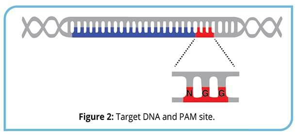
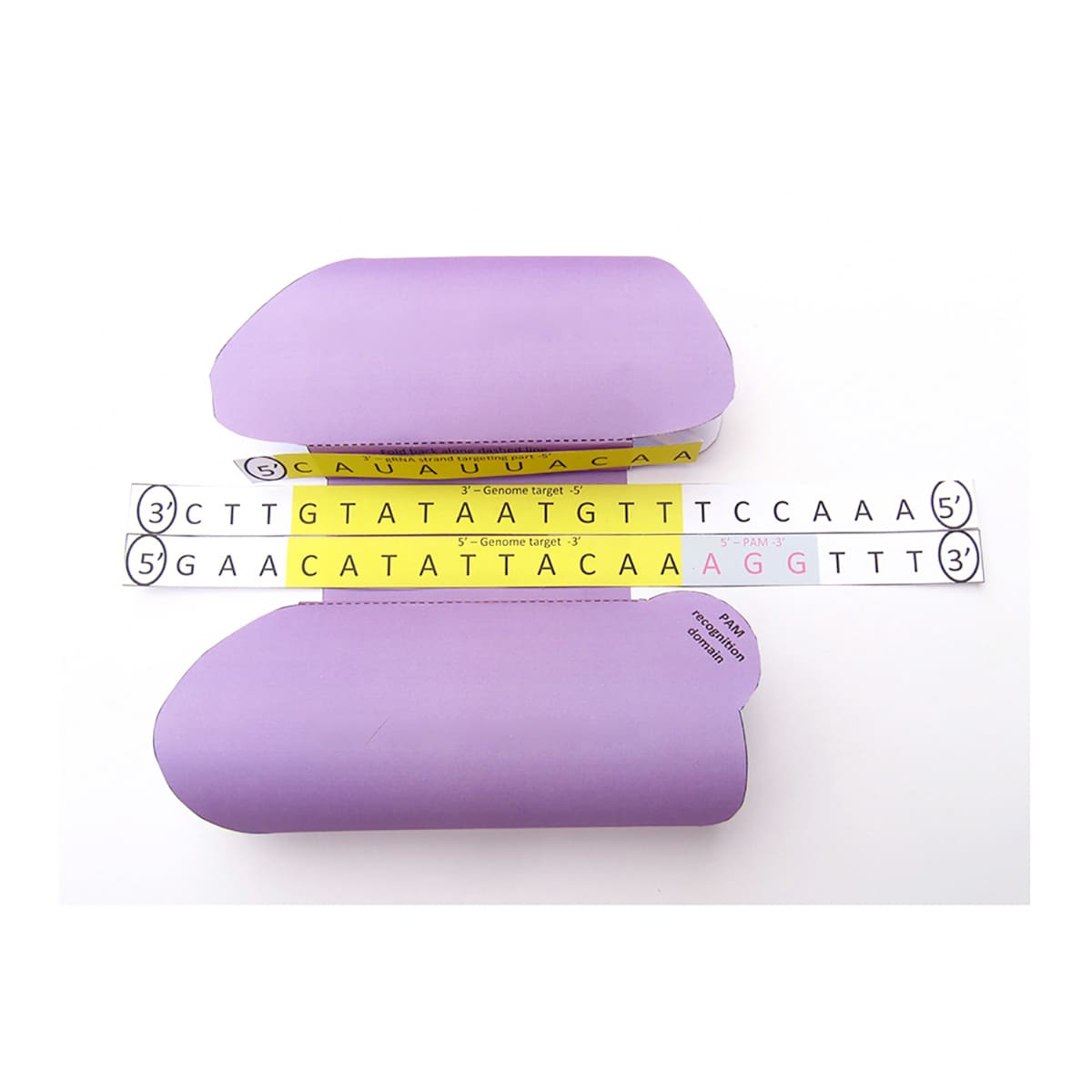
Thanks in part to the newly available DNA sequencing data, a study led by Ruud Hansen found that the Cas enzymes could snip DNA but didn’t know why. At the same time, Alexander Bolotin’s team at the French National Institute for Agricultural Research found that the spacers all share a common sequence they called the protospacer adjacent motif (PAM). The PAM enables Cas enzymes to recognize their target. Different Cas enzymes recognize different PAM sequences; the most commonly-used Cas9 from Streptococcus pyogenes recognizes the PAM sequence 5'-NGG-3', where “N” can be any nucleotide base (Figure 2).
 The discovery that CRISPR spacers were related to viral DNA sequences occurred by three different groups of scientists. Eugene Koonin, an evolutionary biologist at the National Center for Biotechnology Information in Bethesda, Maryland, developed a theory that bacteria were using CRISPR to fight off viruses. Koonin’s theory was tested by Roldolphe Barrangou and Philippe Horvath, then microbiologists at the yogurt company Danisco in France. The company used bacteria to convert milk into yogurt, and entire cultures could be wiped out by bacteria-killing viruses. Barrangou and his team infected one of their yogurt bacteria – Streptococcus thermophilus – with two strains of viruses and cultured the resistant bacteria that survived the assault. Upon examination, they found DNA from the viruses they had used inside CRISPR spacers.
 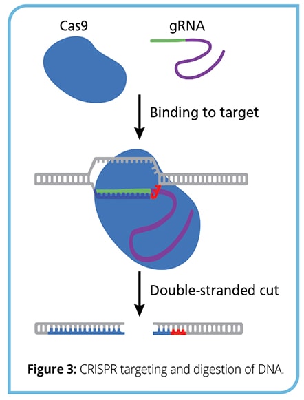 What they discovered was that Cas enzymes could cut DNA and were programmable. Using the CRISPR-Cas9 system from Streptococcus pyogenes, which causes strep throat, Doudna and her colleagues figured out how to hand the Cas9 enzyme an RNA molecule that matched a sequence of DNA they wanted to cut from the genome, then guide it to the target site (Figure 3).
 The ability of CRISPR-Cas to specifically target and cut DNA, combined with modern DNA sequencing, has opened new avenues in genetic engineering, molecular biology, and synthetic biology. Researchers can determine the sequence of a segment of a gene, design a CRISPR guide RNA (gRNA) to specifically cut the DNA, and combine everything within a cell to efficiently change the DNA. The gRNA combines the tracrRNA and CRISPR RNA into a single DNA molecule, simplifying delivery into a cell. One of the most common uses of CRISPR technology is to digest a gene to disrupt its function. Once cut, DNA repair mechanisms will try to mend the double stranded break, often resulting in small insertions, deletions, or other mutations that disrupt gene function.
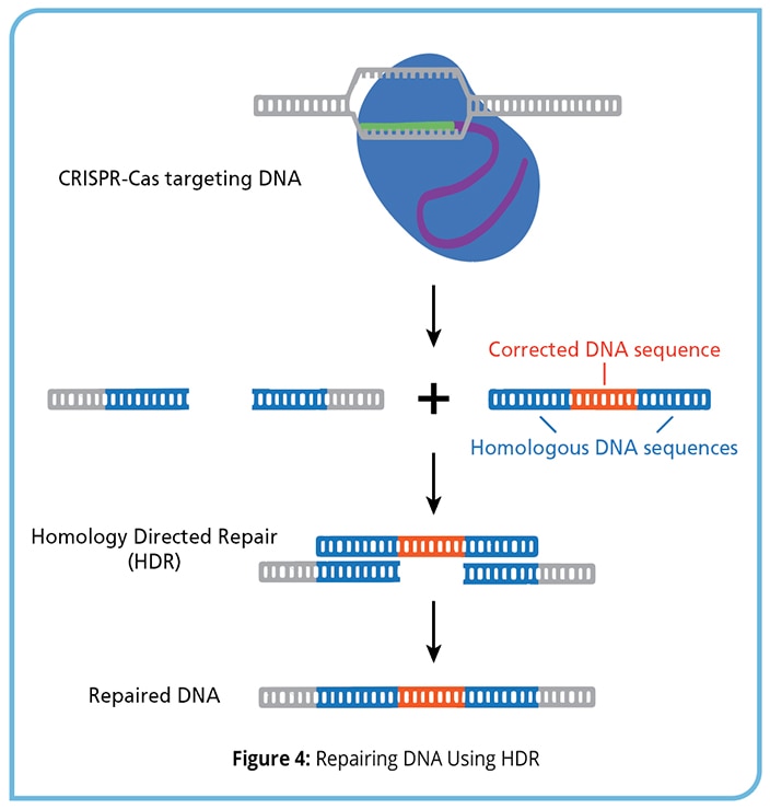 In addition to using CRISPR-Cas systems to disrupt mutated genes, scientists can use CRISPR to replace them with genes that function the way they are supposed to (Figure 4). First, the DNA is cut using CRISPR-Cas to create a double stranded break. Next, the cells are given a template DNA strand, containing the correct sequence, which can be incorporated into the cut DNA using homology directed repair (HDR). With HDR, the natural cellular machinery will incorporate the template DNA into the genome at the site of the CRISPR digest. By controlling the template DNA strand, researchers can repair mutated genes or even insert entirely new genes into an organism. CRISPR-Cas systems allow researchers to easily place the new genes precisely where they want them, unlike some of the older methods of gene therapy where the new genes are randomly inserted into the plant or animal genomes.
 Nature’s creations aren’t formed in a laboratory, they are formed in specific environments for specific purposes and sometimes parts of that original environment are critical to their success. The sickle cell trait is a good example. Sickle cell anemia is an inherited disease caused by a mutation that produces an abnormal hemoglobin protein. The mutated hemoglobin can change the shape of red blood cells, causing them to become rigid and get caught in blood vessels. The sickle cell trait originally developed in Africa as a defense against malaria. The twisted blood cells are resistant to infection from malaria, and cyanate, a chemical found in the local guava and cassava plants, can help to minimize some of the difficulties from the mutated cells. When African people went to parts of the world that did not contain cyanate-rich plants, those oddly shaped red blood cells began to cause additional problems.
Similarly, although initial research has been extremely successful, scientists have discovered a number of unexpected results while using CRISPR-Cas in eukaryotic organisms. For example, although CRISPRCas cleavage is incredibly specific, it is still possible to have off-target effects - sites in the DNA with matching sequences to the guide RNA, as well as unexpected sites that are still targeted and digested. In addition, some studies have linked CRISPR to a potential increase in cancer risk in early non-clinical tests. Therefore, additional experimentation is essential to ensure safety before each round of clinical trials.
The CRISPR mechanism developed in single- celled organisms (bacteria) to fight off viruses. It is possible that our attempts to use this system outside of bacteria is leading to some of these unexpected issues. Scientists are trying to use it in complex, multicellular organisms with thousands of internal wild-card variables and many more environmental variables that come into play.
Basic genetics tells us that, while there are approximately 3 billion base pairs in human DNA, only about 2% of them are organized into genes that can be translated into the messenger RNA (mRNA) that tells our cells how to make proteins. The other 98% of our genome is made up of what we call non-coding DNA, and we have very limited ideas about what that does. So far we have discovered that non-coding DNA plays a role in how genes are expressed, the architecture of the chromosomes, and how we inherit specific traits as a species, but how it does these things is still unclear and there are undoubtedly other functions performed by that mysterious 98% about which we know nothing at all. When we start tinkering with the genome, we can expect surprises, and not all of them will be pleasant ones.
But the only way to find out what we need to know is to begin exploring. It will take years to understand how our genome works and how each part of it affects the others, so we must proceed rigorously and cautiously, a small step at a time. Fortunately, a small step at a time with no object but exploring an interesting phenomenon is a classical description of good science.
Scientists in many countries are now performing hundreds of CRISPR experiments with the diverse goals of repairing defective DNA in mice, editing genes in crops to engineer a better food supply, and rewriting the genome of the elephant to recreate a woolly mammoth. New companies using Doudna, Charpentier, and Zhang’s technologies are starting up to address everything from new cancer treatments to altering insect genomes and eliminating the mosquitoes that carry malaria.
In this experiment, students will simulate the use of CRISPR-Cas9 to target a genetic mutation found in a patient suffering from Cystic Fibrosis. Students will develop an understanding of guide RNA (gRNA) design, and use agarose gel electrophoresis to examine pre-prepared DNA samples after CRISPR treatment.
 Explore cutting-edge biotechnology with this hands-on CRISPR-Cas9 simulation. Students will assume the role of plant geneticists working to develop corn crops capable of surviving, and thriving, in a changing environment. This experiment will allow students to develop an understanding of CRISPR-Cas9 applications in the laboratory, cleave DNA, and examine their results after gel electrophoresis.
 Unlock the exciting world of gene editing and investigate the incredible power of CRISPR technology right from your classroom! In this experiment, students explore cutting-edge genetic engineering using CRISPR-Cas9 to knock out GFP and B-galactosidase genes in classroom safe bacteria. Additionally, students test the specificity of CRISPR-Cas9 first hand by switching the CRISPR RNA templates and analyzing the results.
In this experiment, students will engineer bacteria that are capable of surviving on a distant planet! Students will simulate the use of CRISPR-Cas9 to modify bacterial DNA, which will then be transformed into auxotrophic E. coli that are incapable of surviving on the Martian surface. Only bacteria that receive the successfully edited DNA can survive, thrive, and help to terraform Mars!
 CRISPR is a revolutionary new genetic engineering technique that makes editing genomes easy and inexpensive. It is based on a type of immune system found in many types of prokaryote.