Electrophoresis allows us to separate nucleic acids into bands according to their size, shape, or charge. However, both DNA and RNA are clear and colorless in solution! To analyze our results, we need to be able to see the individual bands created by electrophoresis.
Overview
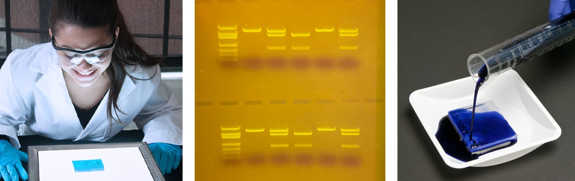
The first DNA visualization techniques used radioactivity or heavy metals to stain the DNA. These techniques were time consuming and required extensive training. Eventually, researchers realized that the fluorescent molecule Ethidium Bromide (EtBr) could be used to stain DNA. This flat, aromatic molecule has a ring structure that mimics a DNA base pair. Molecules of EtBr can slide between the individual base pairs, labeling the DNA. When excited with UV light, the EtBr fluoresces and produces a bright orange light. This technique was easy to perform and very sensitive, allowing researchers to detect very small amounts of DNA.
However, because EtBr is a potential mutagen, it must be handled with care. Furthermore, the UV light necessary to excite the EtBr molecules to fluoresce can cause damage to the eyes and skin. These can be tricky to use in the teaching laboratory! Today, Edvotek offers two simple and effective DNA staining techniques that can be safely used by students. Both produce excellent results, allowing students to visualize the DNA separated by electrophoresis.
What types of DNA stains are available for the classroom laboratory?
Visible dye-based DNA stains are less sensitive than fluorescent stains, but they are an excellent alternative for the teaching classroom because they are non-toxic. The dye molecules stain the DNA fragments a bright blue color that is easily seen by the naked eye, so special equipment is not required to detect the DNA. FlashBlue™ is a visible dye-based DNA stain that offers simple and rapid staining of agarose gels. FlashBlue™ is provided as a concentrated liquid stain that, when diluted, can be used for both rapid and overnight staining of DNA fragments.
Research laboratories commonly use fluorescent DNA stains because they are extremely sensitive, making it easy to detect small amounts of DNA. SYBR® Safe is a DNA stain that fluoresces with a bright green color when excited with light, similarly to EtBr. Unlike EtBr, SYBR® Safe has been engineered to be less mutagenic, making it much safer to use in the classroom. Furthermore, SYBR® Safe can be excited using blue light, which does not have the hazards of UV light.
What do I do after staining the gels FlashBlue™ gels do not need any specialized equipment to visualize the results. The DNA fragments will appear as dark blue bands on a light blue background. However, visualizing the results using a white light transilluminator will help to increase the contrast between the bands and the background. Our White LED Transilluminator features a spacious 25 x 25 cm viewing area illuminated by long life LEDs and is housed in a slim aluminum body. It’s designed to safely enhance the visualization of DNA stained with FlashBlue™ or dye gels.
SYBR® Safe requires UV or blue light to visualize the DNA bands. This is because it is a fluorescent dye that requires bright light to excite the dye molecules that are bound to DNA. When the dye molecules are excited, they emit a bright green light that is easily seen and analyzed. The all-new TruBlu™2 LED Transilluminator utilizes blue light to view DNA gels stained with SYBR® Safe, thus eliminating the need for UV light or ethidium bromide which can be a hazard in the lab. The spacious viewing area measures 27 x 15 cm, which allows you to visualize multiple agarose gels at once. Furthermore, the TruBlu™2 also has a white light feature which is perfect for FlashBlue™ stained gels or dye electrophoresis gels.
 Electrophoresis allows us to separate nucleic acids into bands according to their size, shape, or charge. However, both DNA and RNA are clear and colorless in solution! To analyze our results, we need to be able to see the individual bands created by electrophoresis.
 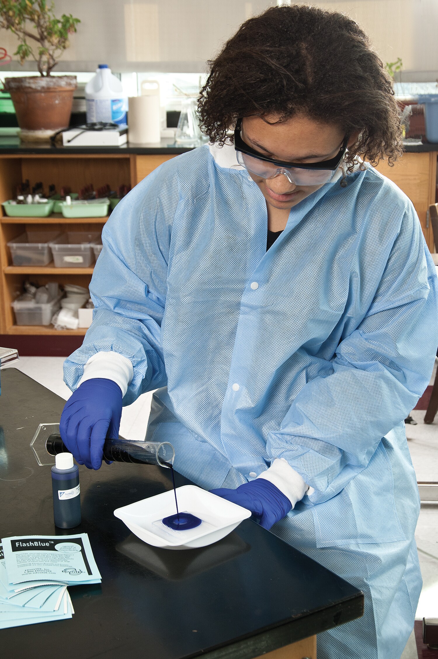 The first DNA visualization techniques used radioactivity or heavy metals to stain the DNA. These techniques were time consuming and required extensive training. Eventually, researchers realized that the fluorescent molecule Ethidium Bromide (EtBr) could be used to stain DNA. This flat, aromatic molecule has a ring structure that mimics a DNA base pair. Molecules of EtBr can slide between the individual base pairs, labeling the DNA. When excited with UV light, the EtBr fluoresces and produces a bright orange light. This technique was easy to perform and very sensitive, allowing researchers to detect very small amounts of DNA.