THE BIOTECHNOLOGY
EDUCATION COMPANY
EDUCATION COMPANY
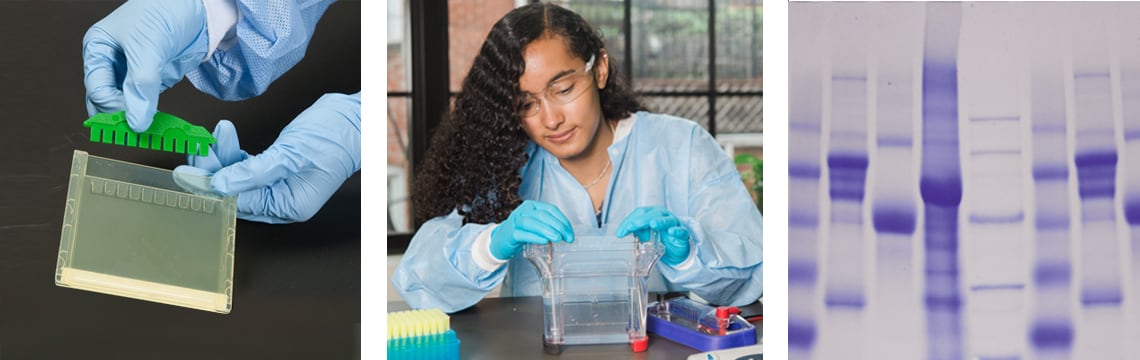 We might just think of protein as something in our daily diet, but proteins are extremely important biological molecules. Each protein is a long chain built from linked amino acids called a polypeptide. Within an organism, proteins are responsible for cell communication, structure, enzyme activity, and more. Through chemical analysis, we know that proteins make up 50% or more of the cell’s total volume. Using biotechnology techniques, we can study these versatile macromolecules in your classroom laboratory.
 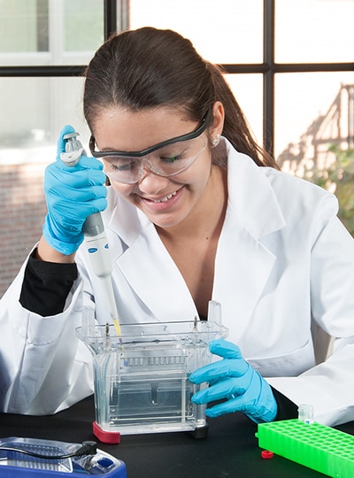 Proteins were first discovered in the 18th century by biologists who were exploring the components of cells. Researchers extracted these molecules and then observed the physical changes that happened when they were added to acid or heat. For example, one of the first proteins identified was albumen, which is present in the clear, viscous liquid that we call the white of the chicken egg. When heated, however, the egg white hardens and changes color to become opaque and white.
 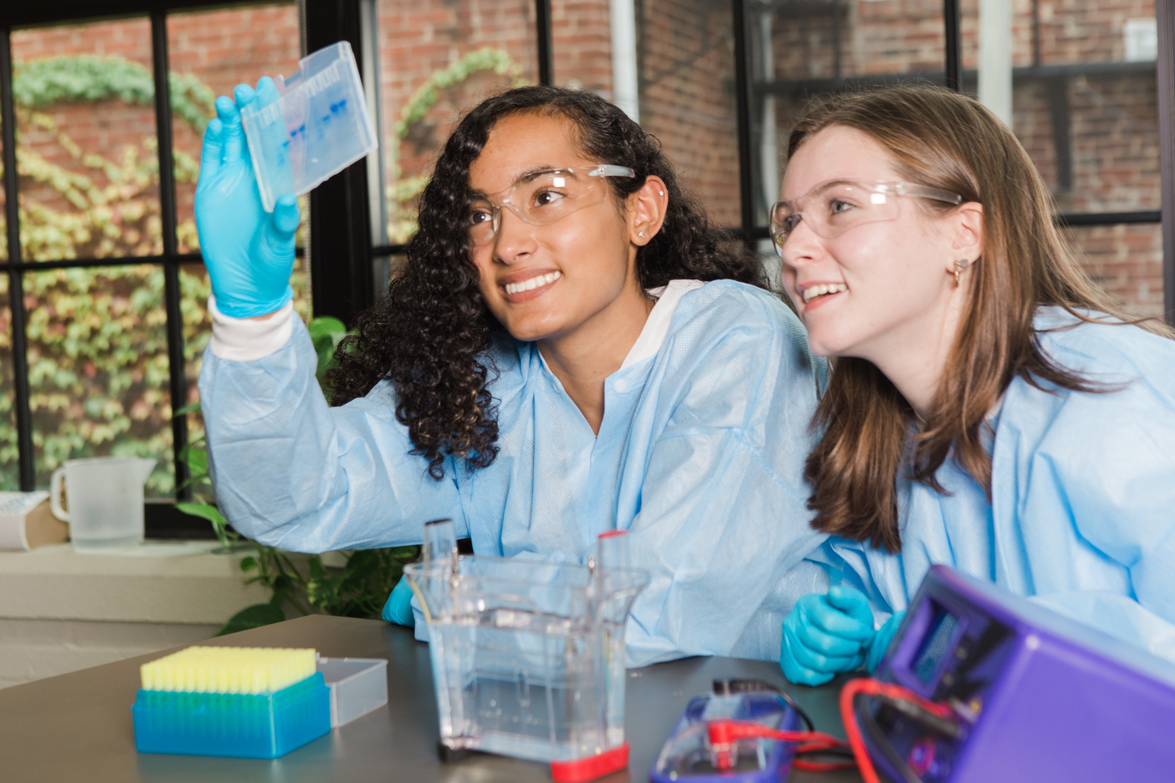 "Edvotek at Home" is a set of resources to teach the basics of Edvotek’s labs through worksheets and presentations. While we believe in the importance of hands-on learning, these free online learning tools are ideal if you can not perform the hands-on experiments in class. Each set includes a student sheet, an instructor’s guide, and an accompanying powerpoint presentation and results sheet. This resource is provided in a downloadable zipped folder below.
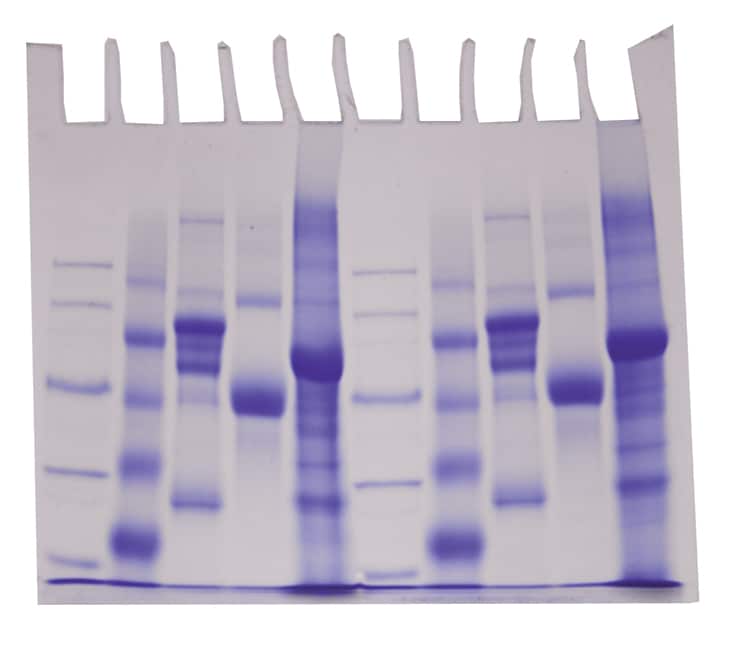
Using Biotechnology to Diagnose HIV/AIDS - The Human Immunodeficiency Virus (HIV) causes acquired immune deficiency syndrome (AIDS), a serious disease that suppresses a patient’s immune system, leaving them susceptible to infections. In this simulation, we’ll perform two common tests (western blot, ELISA) used by doctors to diagnose an HIV infection.
A Bright Idea: Using GFP to Teach STEM - Bring exciting STEM learning techniques into your classroom laboratory! In this hands-on workshop, we will build a size-exclusion chromatography column. The column is used to purify green fluorescent protein (GFP) from a crude bacterial extract. Proteins containing fractions are identified by fluorescence and analyzed for purity by SDS-PAGE.
Troubleshooting Guide - Frequently asked questions, common mistakes, and how to course correct your experiment: